
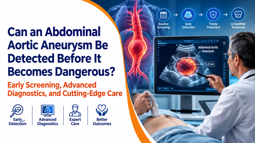
Can an Abdominal Aortic Aneurysm Be Detected Before It Becomes Dangerous? Early Screening, Advanced Diagnostics, and Cutting-Edge Care
By Dr Praveen Chandra, Padma Shri, Chairman, Interventional Cardiology, Medanta – The Medicity, Gurugram
In cardiovascular practice, few presentations are as sobering as those accompanied by complete absence of symptoms.
Consider a representative presentation: an active, asymptomatic man in his late sixties presenting for an elective cardiac workup. Despite an unremarkable physical examination and high functional capacity, an abdominal screening scan identified a dilated aorta, an aneurysm that had developed insidiously over years. It serves as a classic reminder of why structural vascular disease requires deliberate screening rather than reliance on clinical warning signs.
This encapsulates the clinical challenge of abdominal aortic disease. An aneurysm can progress insidiously over years, entirely unannounced, in patients who otherwise demonstrate excellent functional capacity.
The transition from a silent expansion to acute vascular compromise is often sudden. Consequently, establishing precise, early diagnostic pathways before clinical deterioration occurs represents one of the most critical imperatives in modern cardiovascular practice.
The Silent Threat: Why Early Detection Changes Everything
The aorta is the body’s principal artery, carrying oxygenated blood from the heart to every organ system. An abdominal aortic aneurysm occurs when a segment of this vessel, as it passes through the abdomen, weakens and balloons outward, typically defined once its diameter reaches three centimetres or beyond. The danger lies not in the aneurysm’s existence but in its trajectory. As it enlarges, the vessel wall grows progressively thinner and less able to withstand the pressure of each heartbeat, and the risk of eventual rupture rises accordingly.
A ruptured abdominal aortic aneurysm is among the most lethal emergencies in vascular medicine. Evidence reviewed by international preventive health authorities has placed the mortality associated with rupture as high as eighty per cent, a figure that stands in stark contrast to the outcomes of a planned, elective repair performed before rupture occurs. This single statistic explains, more persuasively than any other, why the distinction between proactive detection and emergency intervention is not a matter of clinical preference but one of survival itself.
The reason this condition evades notice is structural to the disease process. Most patients living with an abdominal aortic aneurysm experience no symptoms whatsoever until it ruptures or grows large enough to press against surrounding structures. When symptoms do appear, they are often subtle and easily misattributed: a vague sense of fullness or pulsation in the abdomen, mild back discomfort, or nothing at all. This is not a failure of patient awareness. It is the defining clinical challenge of the disease, and it is precisely why detection cannot reasonably depend on waiting for symptoms to appear.
Who Is at Risk? Clinical Risk Factors and International Screening Guidelines
International guidance from bodies including the Society for Vascular Surgery and the European Society of Cardiology, alongside the widely cited recommendations of the U.S. Preventive Services Task Force, converges on a consistent risk profile for this condition, one worth understanding in some depth rather than treating as a vague statistical footnote.
Age remains the most significant factor, with risk rising notably from the age of sixty-five onward. A history of smoking, even one that ended decades earlier, is among the strongest known contributors, and the combination of age and smoking history forms the basis of the clearest international screening recommendation available: a one-time abdominal ultrasound for men between sixty-five and seventy-five who have ever smoked. Men are affected considerably more often than women, though when women do develop the condition, evidence suggests it tends to progress more aggressively and is more frequently discovered later.
Hypertension, elevated cholesterol, and established atherosclerotic disease elsewhere in the body all compound the risk meaningfully, given that an abdominal aortic aneurysm frequently coexists with broader cardiovascular disease rather than occurring as an isolated finding. Family history carries particular weight as well. A first-degree relative, a parent or sibling, diagnosed with an abdominal aortic aneurysm roughly doubles an individual’s own risk, making a candid family health conversation one of the more valuable, low-cost diagnostic tools available to any patient.
The gold-standard screening method for this population is abdominal ultrasound, a painless, radiation-free examination that takes only about ten minutes to perform. Its diagnostic accuracy is exceptional, with sensitivity and specificity for detecting an aneurysm approaching near-total reliability, which is what makes it such a compelling public health tool. A brief, low-risk scan, performed once in an at-risk individual, can identify a condition that would otherwise remain invisible until the moment it becomes a medical emergency.
Can It Be Detected Early? Advanced Diagnostic Modalities
Once an aneurysm has been identified, or is suspected on clinical grounds, the diagnostic conversation moves beyond a single ultrasound scan into a more nuanced choice of imaging modalities, each suited to a different stage of evaluation.
Abdominal ultrasound remains the appropriate first step for both initial detection and ongoing surveillance. It requires no contrast dye, no radiation exposure, and can be repeated safely and affordably at whatever interval a patient’s risk profile demands, making it ideal for the long-term monitoring of smaller, stable aneurysms.
Contrast-enhanced CT angiography, by contrast, offers a considerably more detailed, three-dimensional view of the aneurysm’s precise anatomy, its relationship to nearby arteries branching toward the kidneys and pelvis, and the exact measurements required for treatment planning. This modality becomes essential once an aneurysm approaches a size or growth rate that may warrant intervention, since the fine anatomical detail it provides directly informs whether a minimally invasive repair is feasible and, if so, precisely how it should be planned.
Magnetic resonance angiography serves as a valuable alternative for patients in whom iodinated contrast dye used in CT imaging is best avoided, such as those with significant kidney impairment, offering comparable anatomical detail through a different imaging principle.
Together, these three modalities allow a vascular and cardiovascular team to move a patient seamlessly from initial, low-cost detection through to the granular, procedure-ready imaging required for safe intervention, without unnecessary radiation or invasive testing at any stage that does not demand it.
Understanding Size Thresholds
The size of an aneurysm, and critically, the rate at which it is growing, determines the appropriate clinical pathway far more than the diagnosis itself. Aneurysms measuring below four centimetres are generally managed through periodic surveillance imaging alone, often at intervals of two to three years, since the annual rupture risk at this size remains low. As an aneurysm approaches four to five centimetres, surveillance typically intensifies to every six to twelve months, allowing the clinical team to track its trajectory closely. Intervention is generally considered once an aneurysm reaches approximately five and a half centimetres in men, a slightly lower threshold in women, or when it demonstrates rapid growth, generally defined as an increase of more than one centimetre within a single year, regardless of its absolute size at that point.
Modern Treatment Paradigms: From Surveillance to EVAR
For the majority of patients diagnosed with a small aneurysm, treatment begins not with a procedure but with a disciplined programme of surveillance and risk factor modification, including blood pressure control, smoking cessation, and management of coexisting cardiovascular disease. This measured, evidence-guided approach, rather than reflexive early surgery, reflects the current international consensus and spares patients the risks of an intervention before it is genuinely warranted.
For aneurysms that do reach a threshold requiring active treatment, the field has been transformed over the past two decades by endovascular aneurysm repair, widely known as EVAR. Rather than the extensive abdominal incision required by traditional open surgery, EVAR is performed through small punctures, typically in the groin, through which a catheter delivers a fabric-covered stent graft to the precise site of the aneurysm. Once deployed, the graft reinforces the weakened vessel wall from within, redirecting blood flow away from the aneurysmal sac and substantially reducing the risk of rupture.
The clinical advantages of this minimally invasive approach are well documented in the international literature: markedly reduced blood loss compared with open repair, a shorter hospital stay, and a meaningfully faster return to normal activity for most patients. The precise choice between an endovascular and a traditional open approach depends on the aneurysm’s anatomy, including its shape, its relationship to nearby arteries, and the overall health profile of the patient, and is best determined through detailed imaging and a candid, individualised discussion between patient and specialist rather than a one-size-fits-all protocol.
Why Choose Gurgaon and Medanta for Aneurysm Care
For patients in India and those travelling from abroad in search of a trusted abdominal specialist for aneurysm care in Gurgaon, the quality of outcome rests on more than the treatment itself. It depends on the completeness of the surrounding clinical ecosystem, from accurate early detection through careful surveillance to access, when needed, to the full range of modern endovascular treatment options, all coordinated under a single, experienced team.
At Medanta, The Medicity, Gurugram, this ecosystem has been built with particular deliberateness. The hospital’s hybrid operating suites allow advanced imaging and catheter-based intervention to take place within the same sterile environment as open surgical capability, meaning that a patient’s precise anatomical needs, rather than departmental boundaries, determine how their care unfolds. Dedicated cardiac catheterisation laboratories, high-resolution CT and MRI imaging, and specialised intensive care units with continuous haemodynamic monitoring support every stage of this pathway, from initial diagnosis through post-procedural recovery. Medanta holds accreditation from the Joint Commission International, alongside national accreditation from NABH and NABL, reflecting adherence to globally benchmarked standards of patient safety and clinical quality.
Our cardiovascular and vascular specialists work in close, continuous collaboration, ensuring that a patient identified with an aneurysm, whether through deliberate screening or an incidental finding during an unrelated evaluation, is guided through every subsequent decision with clinical rigour and genuine continuity of care, rather than being handed between disconnected departments at each stage.
A Seamless Pathway for International Patients
For patients travelling from the Gulf and the wider Middle East, from Africa, from the CIS countries, from elsewhere in South Asia, and from the United Kingdom, the United States, and other Western countries, Medanta’s international patient services are designed to remove the friction that often accompanies care sought far from home. This typically includes initial tele-consultation and remote second-opinion review before travel is even arranged, dedicated support with medical visa documentation, airport transfers, multilingual patient coordinators who remain a consistent point of contact throughout the visit, and transparent, itemised treatment cost estimates provided well in advance. Many international patients also find that the overall cost of equivalent, globally benchmarked endovascular care in Gurgaon compares favourably against comparable treatment in Western healthcare systems, without any compromise in clinical standard or technology. Recovery protocols are similarly structured with the travelling patient in mind, including coordinated discharge planning and virtual follow-up consultation once a patient has returned home, so that the relationship with the treating team does not end at the airport.
Frequently Asked Questions
Is an abdominal aortic aneurysm always fatal if left undetected?
Not necessarily, but the risk compounds silently over time. Many aneurysms remain small and stable for years under appropriate surveillance. The danger arises specifically when a large or rapidly growing aneurysm goes undetected and eventually ruptures, an event associated with very high mortality. This is precisely why early detection, rather than reactive treatment, is emphasised throughout international guidelines.
Is it safe to travel internationally for treatment if I have a diagnosed aneurysm?
In most cases, yes, provided the aneurysm has been properly evaluated and sized beforehand, and the patient has clearance from their treating physician for travel. Aneurysms below the threshold requiring intervention generally do not restrict air travel. For those approaching or exceeding the size threshold for treatment, coordinating evaluation and, where appropriate, treatment before extended travel is the more prudent course, and this is precisely the kind of assessment a pre-travel tele-consultation is designed to clarify.
How long is the recovery after endovascular aneurysm repair?
Recovery following EVAR is considerably shorter than after traditional open surgery. Many patients are mobile within a day of the procedure and can expect a hospital stay of a few days, followed by a gradual return to normal activity over several weeks, though the precise timeline depends on the individual patient’s overall health and the complexity of the repair.
How accurate is ultrasound screening for detecting an aneurysm?
Abdominal ultrasound is highly accurate for this purpose, with sensitivity and specificity that approach near-total reliability in detecting the presence of an aneurysm. It is the internationally recommended first-line tool for both initial screening and long-term surveillance, reserving more detailed imaging such as CT angiography for cases requiring precise anatomical planning ahead of intervention.
At what size does an aneurysm require treatment rather than monitoring?
Intervention is generally considered once an aneurysm reaches approximately five and a half centimetres in men, a somewhat lower threshold in women, or when it grows more than one centimetre within a year regardless of its absolute size. Aneurysms below these thresholds are typically managed through structured surveillance imaging rather than immediate treatment.
There is a certain quiet discipline to preventive vascular medicine that I find genuinely compelling. Unlike the drama of an emergency intervention, detecting an abdominal aortic aneurysm early offers no dramatic moment of rescue, no visible triumph. Its reward is instead an absence, the crisis that never occurs, the rupture that never happens, because the condition was identified and managed years before it had the chance to become an emergency at all.
This, to me, represents medicine at its most quietly powerful. Not the drama of intervention under pressure, but the foresight to look for what has not yet announced itself, and the discipline to act on what is found with patience rather than alarm.
If you have risk factors for abdominal aortic aneurysm, a relevant family history, or wish to discuss whether screening or evaluation is appropriate for you, Dr. Praveen Chandra and the cardiovascular and vascular team at Medanta, Gurugram, are available for consultation. International patients can begin with a tele-consultation and remote second opinion before travel, supported by our dedicated coordination team throughout evaluation, treatment, and recovery. Reach out to schedule an assessment and take a proactive step toward long-term vascular health.
— Dr. Praveen Chandra