
What Happens Inside the Cath Lab During Emergency Heart Attack Treatment
Dr. Praveen Chandra
Chairman, Interventional & Structural Heart Cardiology, Medanta
Padma Shri (2016) | Performed India’s First TAVI | Pioneer of Structural Heart Intervention in India
In the case of a heart attack, every minute is important in saving the amount of heart muscle that can still be salvaged. Cardiology has undergone many changes throughout its development. A few decades ago, treatment options were basically symptom relief and complication prevention. Nowadays, we are able to clear a clogged artery quickly through minimally invasive methods using a catheter. This development has saved countless lives around the world and helped them avoid major disabilities.
For patients and their loved ones, the process of being moved from the emergency department to the cath lab is unknown and frightening.
Why Immediate Treatment Matters
The occurrence of a heart attack, known scientifically as an acute myocardial infarction, happens due to the sudden blocking of a coronary artery, which is responsible for providing oxygenated blood to the heart. In most instances, the blockage happens due to the rupture of cholesterol plaque in the artery, which leads to the formation of a blood clot. As a result, the part of the heart supplied with oxygen is damaged irreversibly because there is no blood supply.
There exists a phrase used by cardiologists, which goes, “Time is muscle.” In other words, each passing minute means the death of more heart muscle. Consequently, the goal of the emergency procedure becomes clear: restoration of the blood supply needs to be done as fast and safely as possible.
According to current international guidelines, the door-to-balloon time should be less than 90 minutes for STEMI patients. Many studies have shown that the faster the process of restoring the blood flow was, the less mortality and complications were observed.
The Journey Begins in the Emergency Department
The emergency care of heart attacks begins even before the patient reaches the catheterization laboratory.
After reaching the hospital, doctors quickly analyze symptoms like severe chest pain, shortness of breath, sweating, nausea, or chest pain radiating to the arms, jaw, and back. The electrocardiogram (ECG) test is immediately conducted to detect any specific abnormalities indicating a heart attack.
Blood samples are taken to analyze cardiac markers like troponin, and at the same time, intravenous access is established to provide medication to the patient. Antiplatelets and anticoagulants, along with other medications, may be administered based on the status of the patient.
After the confirmation of a heart attack that requires emergency angioplasty, the team from the cath lab is immediately summoned. Each member of the team realizes the importance of avoiding any delays for survival.
Inside the Cath Lab
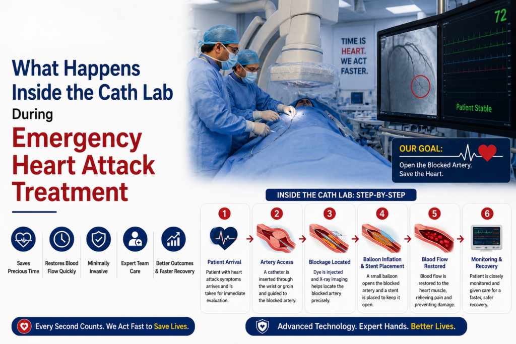
The Cardiac Catheterization Lab is a very special setup where state-of-the-art imaging machines are used to help doctors see inside the coronary arteries on a live basis.
Even though the technology used here is cutting-edge, most people entering this room will be taken aback by how different it is from a traditional operating theater, since there are large screens where X-ray images can be seen,n and also specialized equipment that helps maneuver the devices within the blood vessels of the body.
There is usually an interventional cardiologist, specialist nurses, cardiac technologists, radiographers, anesthetists if required, and other specialists ready to tackle unexpected complications.
In spite of being calm and systematic, everyone working here coordinates well.
Preparing the Patient
Unlike traditional heart surgery, emergency coronary intervention usually does not require opening the chest or placing the patient under general anesthesia.
Most procedures are performed under local anesthesia with light sedation. The patient remains awake and can communicate with the medical team throughout the intervention, although medication helps reduce discomfort and anxiety.
The wrist (radial artery) has become the preferred access site for many procedures because it lowers bleeding complications and allows patients to recover more quickly. In certain situations, particularly when anatomy or clinical circumstances require it, the femoral artery in the groin may be used instead.
After cleaning and sterilizing the access site, a small sheath is inserted into the artery. Through this sheath, the interventional cardiologist introduces slender catheters that travel through the arterial system toward the heart under continuous X-ray guidance.
To the patient, the procedure is generally painless. Most people feel only the initial injection of local anesthetic and occasional sensations of warmth when contrast dye is injected.
Identifying the Blocked Artery
The very first step is to do a coronary angiography.
A contrast agent is infused via the catheter as high-resolution fluoroscopy images are taken that give details of the location and severity of the blockage in the coronary arteries. This also shows if any narrower segments may be involved.
The fact that the cardiologist can see the coronary circulation in real time makes it possible for him to decide the treatment according to the specific anatomy of the patient. Some blockages may be easy while some complex.
The first thing to do after pinpointing the culprit artery causing the heart attack is to open up the blockage.
Restoring Blood Flow Through Primary Angioplasty
Emergency angioplasty, also known as primary percutaneous coronary intervention (PCI), remains the preferred treatment for most patients with STEMI when performed by experienced teams within recommended timeframes.
A very thin guidewire is carefully advanced across the blocked segment of the artery. This serves as a pathway for balloons and other treatment devices.
In many cases, a tiny balloon is first inflated to compress the obstructing clot and plaque against the artery wall. This immediately improves blood flow through the previously blocked vessel.
To provide durable support and reduce the risk of the artery narrowing again, a drug-eluting stent is usually implanted. The stent acts as a small metallic scaffold that keeps the artery open while gradually releasing medication that minimizes excessive tissue growth inside the vessel.
Within moments of restoring circulation, many patients experience significant relief from chest pain. On the imaging monitors, the return of normal blood flow is often visible immediately, a gratifying milestone for the entire treatment team and an encouraging sign for the patient’s recovery.
Beyond Opening the Artery
However, restoration of the blood flow is the main aim of the operation, but the surgery does not necessarily stop after opening the artery.
According to the patient’s status, an interventional cardiologist might choose to employ more advanced intracoronary imaging modalities, namely Intravascular Ultrasound (IVUS) and/or Optical Coherence Tomography (OCT). This way, it becomes possible to take highly accurate images inside the artery to study the plaque’s features, to make sure that the stent is deployed properly, and that its position is the best one.
In some particular cases, it becomes necessary to conduct certain physiological measurements to assess whether any other narrowed vessels need surgical intervention right away, or if they could wait for later treatment.
Monitoring Throughout the Procedure
Despite the fact that angioplasty is minimally invasive, it is conducted under continuous observation.
During the entire procedure, the heart rate, blood pressure, oxygen saturation, and general cardiovascular status of the patient are continuously monitored. There are dedicated nurses, technologists, and doctors working in unison, making sure that any changes in the state of the patient are promptly identified and dealt with.
The duration of most procedures ranges from 30 to 90 minutes; however, in some cases, when the patient suffers from complicated anatomy of the coronary arteries or several blockages, more time is needed.
After the Procedure
Once blood flow has been restored and the intervention is complete, the catheters are removed, and the access site is carefully sealed.
Patients are then transferred to a coronary care unit or intensive cardiac care unit for close observation. During the first 24 to 48 hours, the medical team monitors heart rhythm, blood pressure, kidney function, and signs of recovery while adjusting medications that help prevent future cardiac events.
Many patients notice a significant improvement in chest discomfort and breathing soon after the blocked artery has been opened. However, recovery depends not only on the success of the procedure but also on how quickly treatment is initiated after symptoms begin. Earlier intervention generally preserves more heart muscle and leads to better long-term heart function.
Before discharge, patients receive comprehensive guidance regarding medications, lifestyle modification, cardiac rehabilitation, cholesterol management, diabetes control, smoking cessation, where applicable, and the importance of regular follow-up care.
The Importance of Teamwork
Effective emergency care for heart attacks can never be attributed to one person alone.
Each successful primary angioplasty process involves teamwork among a multidisciplinary team of specialists, including emergency physicians, paramedics, nurses, cath-lab technologists, anesthesiologists when necessary, cardiac intensivists, rehabilitation therapists, and interventional cardiologists.
This expertise ensures that patients can have evidence-based care at each step of the emergency process.
The current management of heart attacks has developed into a scenario where preparation and experience become just as critical as the technology involved.
An Honest Perspective
Primary angioplasty has become the preferred treatment for many acute heart attacks because it consistently achieves higher rates of artery reopening and lower rates of recurrent heart attack and stroke compared with clot-dissolving medications when performed promptly by experienced teams.
However, no medical procedure is entirely without risk.
Bleeding, damage to blood vessels, allergic reactions to contrast dye, kidney complications, abnormal heart rhythms, stroke, or the need for emergency surgery are uncommon but recognized possibilities. Fortunately, serious complications are relatively rare, and advances in equipment, imaging, and procedural techniques have continued to improve safety over the past two decades.
Equally important is recognizing that not every patient presents in the same way. Individuals with delayed presentation, cardiogenic shock, multiple blocked arteries, or significant underlying medical conditions often require highly individualized treatment strategies. The best outcomes are achieved when treatment decisions are guided by careful clinical judgment rather than a one-size-fits-all approach.
Why Early Recognition Saves Lives
One of the biggest problems that is seen in heart attack treatment is the delay in seeking treatment.
Most patients brush off their chest pain, thinking it’s nothing but acidity or digestion problems. Others prefer to wait, thinking their condition will improve without any external help. However, such delays lead to additional damage to the heart tissues.
Any sort of chest pressure, pain that goes to the arm, neck, jaw, or back, sudden breathlessness, heavy sweating, nausea, weakness, or fainting are all reasons enough for emergency medical attention.
Frequently Asked Questions
Is emergency angioplasty painful?
Most patients experience very little discomfort during the procedure. Local anesthesia numbs the access site, and mild sedation helps patients remain comfortable throughout the intervention.
How long does a patient stay in the hospital after a heart attack?
Hospital stay varies depending on the severity of the heart attack and the patient’s recovery. Many uncomplicated cases are discharged within two to four days, while more complex situations may require longer observation.
Will a stent cure heart disease permanently?
A stent treats the blocked artery responsible for the heart attack, but it does not eliminate the underlying process of coronary artery disease. Long-term success depends on medications, healthy lifestyle choices, regular exercise, cholesterol control, diabetes management, and follow-up care.
Can patients return to normal life after emergency angioplasty?
Many patients resume normal daily activities within a few weeks, although recovery timelines vary. Participation in a structured cardiac rehabilitation program significantly improves physical recovery, confidence, and long-term cardiovascular health.
One of the best things about the current state of cardiology is that having a heart attack is no longer considered something beyond help. Modern emergency coronary interventions have helped countless patients, assuming that they start being administered as soon as possible.
The cardiac catheterization lab is the place where experience, technology, and collaboration combine for one goal: restoration of the blood supply before the damage becomes irreversible. Every success story serves as a confirmation of the importance of prompt action and the right treatment.
In case you or anyone around you starts feeling the symptoms of a heart attack, do not let time pass by. You should call for emergency help as soon as possible.
— Dr. Praveen Chandra

