By Dr Praveen Chandra | Padma Shri | Chairman, Interventional Cardiology, Medanta — The Medicity | Performed India’s first TAVI, 2010
You cannot cure what you can only glimpse in the dark. For most of the history of cardiology, we relied on angiograms, grainy, moving shadows to decide how to treat a blocked artery. When patients pointed to the screen and asked, “Is that it?” we were often guessing at what those shadows concealed. Today, modern angioplasty has evolved. We have stopped guessing at the shadow and started measuring the reality. That is what we mean by Precision PCI.
First, What PCI Actually Is
PCI stands for percutaneous coronary intervention, the formal term for what most people call angioplasty. When a coronary artery, one of the small vessels that feed the heart muscle itself, becomes narrowed by plaque, we can reopen it without surgery. We pass a fine tube from an artery in the wrist or groin, thread a soft wire across the narrowing, widen it with a small balloon, and in almost every case leave behind a stent, a tiny scaffold that holds the vessel open. It is among the most common procedures in all of medicine, and it has saved countless lives, including in the middle of a heart attack. Precision PCI uses that very same procedure, performed with far better eyes and a way to measure what truly matters.
The Limits of the Shadow
When we look at a standard angiogram, we are looking at a riverbed, not the riverbanks. The dye shows us where blood flows, the lumen, but it completely blinds us to the artery wall itself.
As interventionists, we must teach the next generation to look beyond the silhouette. An angiogram carries massive blind spots: it hides the plaque burden within the walls, masks calcification, and often misleads us about the vessel’s true size. We might look at a lesion and think it’s critical, when physiologically, the flow is perfectly fine. Conversely, we might place a stent and assume it’s perfect, while the shadow conceals an under-expanded frame that risks thrombosis.
Relying solely on the angiogram is practising yesterday’s medicine. Precision PCI means using advanced imaging and physiology to eliminate the blind spots that shadows leave behind.
What Precision PCI Actually Is
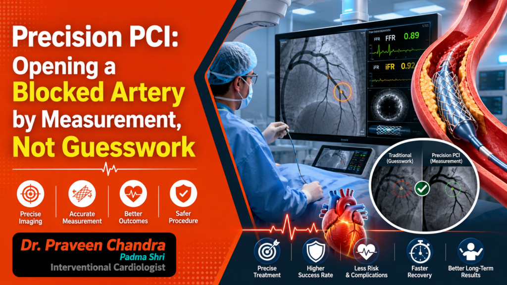
Precision PCI adds two things the angiogram cannot give us, and together they change the quality of the work. The first is intravascular imaging. We pass a probe slimmer than the artery itself, using either sound, which we call IVUS, or light, which we call OCT, and we look at the vessel from the inside. All at once, we can see the wall, the plaque, the calcium, and the exact width of the vessel, which tells us the precise size and length of stent it needs. The second is physiology. Using a hair-thin pressure wire passed gently across a narrowing, a measurement known as FFR, we can tell whether that narrowing is genuinely starving the heart muscle of blood, or whether it merely looks dramatic on the screen. One tool lets us see properly. The other lets us measure properly. Between them, guesswork gives way to fact.
The Procedure, From Start to Finish
Let me walk you through a Precision PCI as the patient lives it. You are awake, lightly sedated, and comfortable. We begin almost always through the wrist, which is gentler and lets you sit up soon afterwards. A fine catheter is guided to the mouth of the coronary arteries, the dye is given, and the familiar angiogram appears on the screen. Here is where the approach parts ways with the old habit. Rather than deciding from the picture alone, we ask two questions in turn. First, does this narrowing actually matter, which the pressure wire answers, so that we treat the blockages truly starving the muscle and leave the innocent ones untouched. Second, if it does matter, what exactly does this vessel need, which the imaging answers by showing us its real size and the nature of the plaque within it. Armed with these exact dimensions, we finally widen the artery and deploy the stent. We are no longer eyeballing the lesion; we are choosing the precise size and length based on mathematical certainty rather than visual estimation. What follows is a step I treat as an absolute mandate. We image the artery once more from within, confirming beyond a shadow of a doubt that the stent is fully expanded and perfectly conformed to the vessel wall along its entire length. A stent that is even slightly under-expanded is the single greatest reason stents fail in the years that follow, and the camera catches what the shadow would have quietly missed. The patient feels none of this beyond a little pressure. The whole difference lives in the detail.
Why It Is Better, and Safer
The evidence has caught up with the logic. In patients with complex disease, guiding the procedure with intravascular imaging has been shown to lower the combined risk of cardiac death, heart attack, and the need to return and reopen the same vessel, set against the angiogram alone. The reason is plain. A stent chosen by measurement and confirmed fully open fails less often and clots less often. The pressure wire, for its part, spares many patients a stent they never needed, which is its own quiet form of safety. There is something here worth knowing as a patient. The reality is that this level of precision is not yet universal. Across the country, a vast number of angioplasties are still guided solely by the traditional angiogram, frequently due to the constraints of time, resource limitations, or cost. But as a field, we must bridge this gap. It is entirely reasonable, before a planned procedure, to ask your cardiologist whether intravascular imaging and pressure measurement will be used to guide your stent. A thoughtful answer is a sign that you are in careful hands.
Why This Is How We Work at Medanta
I have spent much of my career in the coronary arteries, through the long evolution of angioplasty from a blunt instrument into the precise discipline it has become. At Medanta, Precision PCI is how we prefer to work as a matter of course, rather than a flourish reserved for the difficult cases, because we would rather treat the artery we can measure than the one we can only imagine. The exact same philosophy that governs our structural heart interventions, where absolutely nothing is left to assumption, guides our hands in the coronary arteries. In this department, precision is not an occasional choice; it is a daily habit. It is proven by the rigorous pressure measurements we take before we ever commit to a stent, and it is validated by the definitive imaging we perform afterwards to guarantee that the stent is perfectly deployed before the patient ever leaves our care.
If you or someone you love is facing an angioplasty, look your cardiologist in the eye and ask the one question that matters most: ‘Will this be guided by measurement, or just by the shadow?’ And remember, you shouldn’t wait for an emergency to listen to your body. If exertion has begun to bring on chest tightness or breathlessness, seek expert care now, before the artery forces the issue. The heart always rewards those who listen to it early.
“Will this be guided by measurement, or just by the shadow? The heart always rewards those who listen to it early.”

