
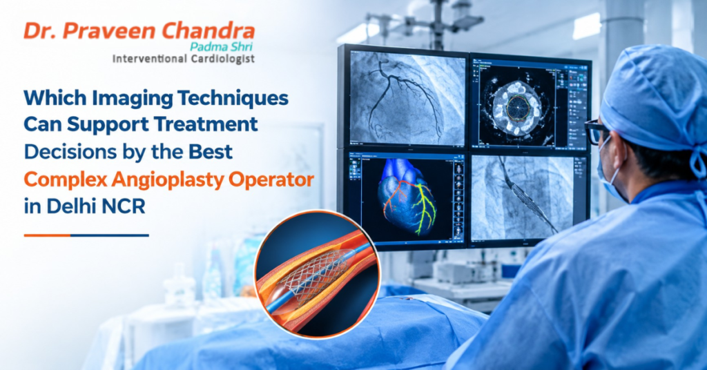
A coronary angiogram has long been the standard way to see blockages in the heart’s arteries. But it has limits. It shows the outline of the blood channel, not the wall of the artery, and it cannot always tell how much a blockage is actually restricting blood flow. In complex cases, such as multiple blockages, heavy calcium, left main disease or chronic total occlusions, additional imaging often makes the difference between a well-judged treatment and a guess.
Patients searching for the best complex angioplasty operator in Delhi NCR may not realise how much of a good result depends on what is seen before and during the procedure. This guide explains the main imaging and measurement tools, what each one shows, and when it is typically used.
Key takeaways
• An angiogram alone shows only the inside outline of the artery. It does not show the artery wall or blood-flow impact.
• Intravascular imaging (IVUS or OCT) shows the artery from the inside, and helps choose stent size and confirm that it is well expanded.
• Pressure measurements (FFR or iFR) show whether a blockage truly restricts blood flow.
• CT and MRI help with planning, and with deciding whether heart muscle can recover.
• The right tool depends on the case. More imaging is not always better.
The main tools at a glance
Technique | What it shows | Typical use in complex angioplasty | Limitations |
Coronary angiography | Outline of the artery’s channel using contrast dye and X-rays. | Basic map of blockages, and guidance during the procedure. | Does not show the vessel wall, plaque type or blood-flow impact. |
IVUS (intravascular ultrasound) | Cross-section images from inside the artery using ultrasound. | Measuring vessel size, assessing plaque and calcium, choosing stent size, and checking expansion. Valuable in left main and long lesions. | Lower detail on the surface than OCT. Needs an extra catheter. |
OCT (optical coherence tomography) | Very high-resolution images using near-infrared light. | Detailed look at plaque, calcium thickness and stent edges and apposition. | Needs contrast to clear blood. Penetrates less deeply, and is less suited to very large vessels or certain ostial lesions. |
FFR / iFR (pressure wire tests) | Whether a blockage is significantly limiting blood flow. | Deciding which blockages need stenting, especially in multi-vessel disease. | A physiology test rather than an image. Not suitable in every situation. |
Coronary CT angiography | 3D images of the arteries, calcium and plaque, without entering the artery. | Planning, especially for chronic total occlusions, calcium assessment and bypass graft evaluation. | Image quality drops with heavy calcium, fast heart rate or irregular rhythm. Uses contrast and radiation. |
Echocardiography | Heart pumping function, wall movement and valves. | Baseline heart function and risk assessment. | Does not show coronary arteries directly. |
Cardiac MRI / nuclear tests | Heart muscle blood supply and viability (whether muscle can recover). | Deciding whether opening a blocked artery is likely to help. | Less widely available, and not needed in every patient. |
How each tool supports decisions
Angiography: the starting map
Does not show coronary arteries directly. Angiography remains the basic roadmap. The operator injects contrast dye into the coronary arteries and watches it with X-rays. In complex disease, the angiogram is combined with other tools because it cannot show everything.
IVUS and OCT: seeing from the inside
Intravascular imaging uses a tiny imaging catheter passed inside the artery. It helps the operator to:
• Measure the true size of the artery so the stent is not too small or too large
• See how much calcium is present and where
• Identify the type and extent of plaque
• Check after stenting that the stent is fully expanded and well pressed against the wall
• Spot problems such as a tear at the stent edge that might otherwise be missed
Guidelines support the use of intravascular imaging in complex situations, such as left main disease and complex stenting. IVUS and OCT each have strengths. IVUS sees deeper into the artery wall and is generally easier to use in large vessels, while OCT gives sharper detail but requires clearing blood with contrast dye.
FFR and iFR: does this blockage matter?
A blockage may look severe on an angiogram yet not limit blood flow enough to cause problems, or the reverse. A thin pressure wire passed across the blockage measures the difference in pressure before and after it. This gives a number that helps decide whether a stent is needed. In patients with several blockages, this can avoid unnecessary stenting and focus treatment on the lesions that matter.
CT angiography: planning from outside the artery
A coronary CT scan can show the arteries, calcium and plaque before the procedure. It is especially helpful when planning chronic total occlusion procedures, where understanding the course and length of the blocked segment is important, and for assessing bypass grafts.
Echocardiography, MRI and nuclear imaging: heart function and viability
An echocardiogram shows how well the heart pumps and whether valves are working normally, which affects both risk and the choice between angioplasty and surgery. Cardiac MRI or nuclear imaging can show whether the heart muscle supplied by a blocked artery is still alive and likely to benefit from reopening it.
Safety: contrast dye, radiation and kidneys
Several of these techniques use contrast dye, X-rays or both. Operators balance the value of extra information against these costs, especially in patients with kidney disease. Strategies include limiting dye volume, using imaging that needs little or no contrast when appropriate, hydrating the patient, and tracking radiation dose. Tell your cardiologist about any kidney problems, diabetes or allergies to dye before your procedure.
Why imaging helps patients
• More accurate decisions: which blockages to treat, and which to leave alone.
• Better stent sizing and placement: avoiding stents that are too small, not fully expanded or poorly positioned.
• Safer handling of calcium: choosing the right preparation tools.
• Fewer surprises: detecting problems during the procedure rather than afterward.
Questions to ask your angioplasty team
• Will you use anything beyond a standard angiogram to decide on stenting?
• Do I need a blood-flow (FFR/iFR) test or imaging inside the artery?
• Would a CT scan or MRI help plan my procedure?
• How will my kidneys be protected from contrast dye?
• How will you confirm the stent is working well afterward?
About Dr. Praveen Chandra
Dr. Praveen Chandra is Chairman of Interventional Cardiology at Medanta Medicity, Gurgaon. He was awarded the Padma Shri by the President of India in 2016 and performed the first percutaneous aortic valve implantation without surgery in India. He has authored over 500 articles and is a reviewer for leading medical journals. His work includes structural heart interventions such as percutaneous tricuspid valve procedures and leadless pacemakers.
When should you see a cardiologist?
Consult a cardiologist promptly if you have chest pain or pressure on exertion, breathlessness, unusual tiredness, or if you have been told that your coronary arteries have significant blockages. Seek emergency care immediately for severe or persistent chest pain, pain spreading to the arm or jaw, sudden breathlessness, or fainting.
Frequently asked questions
What is the difference between IVUS and OCT?
Both create images from inside a coronary artery. IVUS uses ultrasound and sees deeper into the vessel wall, while OCT uses light and gives sharper detail of the inner surface but needs the artery to be cleared of blood with contrast dye. The choice depends on the lesion and the patient.
What is FFR and why is it measured?
Fractional flow reserve (FFR) is a pressure measurement taken across a blockage with a thin wire. It shows whether the blockage is significantly limiting blood flow, which helps decide whether a stent is needed.
Is an angiogram enough to decide on a stent?
An angiogram shows the outline of the artery but not the vessel wall or how much a blockage limits blood flow. In complex cases, additional imaging or pressure measurements often make the decision more accurate.
Can imaging be used if I have kidney disease?
Yes, with planning. Some techniques need little or no contrast dye, and the team can limit dye use and take protective steps for the kidneys. Tell your doctor about any kidney problems before the procedure.
How do I book a consultation for a complex coronary problem?
You can book an appointment with Dr. Praveen Chandra’s team by calling +91 99588 72855 or +91 97172 89363.