
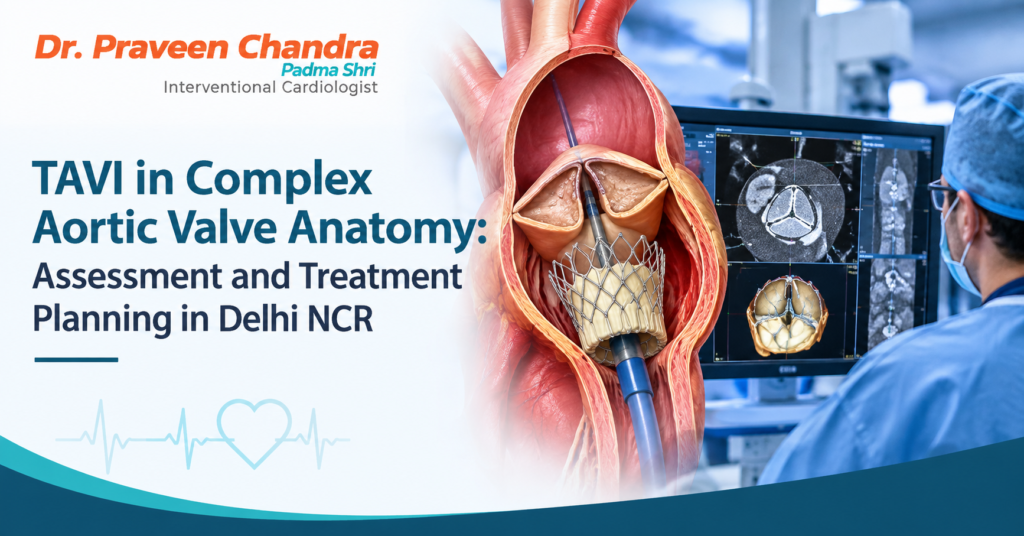
Transcatheter Aortic Valve Implantation (TAVI) is a minimally invasive treatment option for selected patients with severe aortic stenosis. However, factors such as heavy valve calcification, a bicuspid aortic valve, low coronary artery openings and narrow blood vessels can make treatment planning more complex.
Choosing an appropriate TAVI approach requires detailed cardiac imaging, careful valve selection and an assessment of the patient’s overall health. For patients exploring TAVI treatment in Delhi NCR, understanding how specialists evaluate these challenges can help them make informed decisions about their care.
What Makes TAVI Challenging in Complex Valve Anatomy?
TAVI involves delivering a replacement aortic valve through a catheter, commonly via an artery in the groin. It avoids the need for a traditional open-heart incision, although it remains a major procedure with risks that need careful assessment. Certain anatomical features can make planning and valve implantation more complex.
Heavy valve calcification: Calcium deposits can affect how the replacement valve expands and seals against the surrounding tissue.
Bicuspid aortic valve: A bicuspid valve has two leaflets rather than the usual three, which can create a different valve shape and affect planning.
Low coronary artery openings: The position of the coronary arteries may increase the risk of obstruction during or after valve implantation.
Narrow or tortuous blood vessels: Blood vessels that are narrow, calcified or unusually curved may make it difficult to deliver the valve through the usual access route.
Unusual aortic root measurements: The dimensions of the aortic root and surrounding structures help guide valve selection and procedural planning.
These features do not automatically rule out TAVI. A specialist heart team must assess the individual anatomy, likely benefits and risks, and whether TAVI or another treatment would be more appropriate.
How Specialists Assess Valve Anatomy Before TAVI
Cardiac CT and Echocardiography
Cardiac CT is an important part of TAVI planning. It helps clinicians assess the aortic valve, aortic root, coronary artery openings and blood vessels used to deliver the replacement valve. Measurements and the pattern of calcium deposits help the team evaluate valve size, positioning and potential procedural risks.
Echocardiography provides information about valve function, blood flow, pressure differences across the valve and the heart’s pumping ability. Together, these investigations help clinicians plan treatment around the patient’s anatomy rather than relying on standard measurements alone.
Assessing Overall Health
Anatomy is only one part of the assessment. The team may also consider kidney function, other heart conditions, frailty, previous cardiac procedures and the patient’s overall health. For people with impaired kidney function, the use of contrast during imaging may require particular care. The risks associated with significant calcification also need individual evaluation.
How Valve Selection Helps Manage Complex TAVI Cases
Transcatheter valves differ in design, size and delivery system. The choice depends on the patient’s anatomy, calcium distribution, vascular access and other clinical factors. The treatment team weighs these details when deciding which valve and procedural approach are most suitable.
Managing Heavy Calcium Deposits
Extensive or uneven calcification can affect valve expansion and sealing, potentially increasing the risk of paravalvular leakage, where blood flows around the implanted valve. Calcium distribution can also affect nearby structures. Reviewing the imaging helps the team plan implantation and consider the risks relevant to the individual patient.
Assessing a Bicuspid Aortic Valve
A bicuspid aortic valve may have an asymmetrical shape and can be associated with changes in the aorta. CT imaging helps clinicians assess valve dimensions, calcium burden and surrounding structures to determine whether TAVI is appropriate and which replacement valve may be considered. Not every patient with a bicuspid valve is suitable for TAVI; the decision requires individual assessment.
Planning Valve Placement and Reducing Procedural Risks
Accurate positioning is important for valve function. In complex anatomy, detailed imaging and procedural planning help the team anticipate potential challenges and evaluate the result during and after implantation.
Reducing the Risk of Coronary Artery Obstruction
In some patients, the implanted valve or displaced tissue from the original valve can obstruct blood flow into a coronary artery. The team assesses factors such as coronary artery height, aortic root dimensions and the position of the native valve leaflets. If imaging suggests a higher risk, the team may consider protective strategies or a different treatment approach.
Checking for Valve Leakage
The replacement valve should seal appropriately within the native valve structure. Valve selection, positioning and assessment of valve function help the team identify and manage leakage. The result is evaluated during and after the procedure.
What If the Usual Access Route Is Not Suitable?
TAVI is commonly performed through the femoral artery in the groin. If these arteries are too narrow, heavily calcified or tortuous, the team may assess alternative access routes. The choice depends on the patient’s vascular anatomy, overall health, procedural risks and the experience and resources of the treating centre. An alternative route is not automatically safer or more appropriate; the decision must be individualised.
Why a Multidisciplinary Heart Team Matters
Complex TAVI decisions may involve an interventional cardiologist, cardiac surgeon, cardiac imaging specialist, anaesthetist and other clinicians as needed. The team reviews the imaging, other medical conditions, likely benefits and risks, and available treatment options. Depending on the individual case, TAVI or surgical aortic valve replacement may be recommended.
Recovery and Follow-Up After TAVI
After valve implantation, the clinical team monitors recovery and checks for possible complications. Monitoring may include heart rhythm, blood pressure, kidney function and the catheter insertion site. Echocardiography may be used to assess the function of the replacement valve and check for leakage. Recovery varies according to the patient’s health, the complexity of the procedure and whether complications occur.
Follow-up appointments help clinicians assess symptoms, review medicines and monitor valve function. Patients should follow the care team’s instructions and promptly report concerning symptoms such as new or worsening breathlessness, chest pain, fainting or swelling.
How to Choose a TAVI Specialist in Delhi NCR
When discussing treatment, patients can ask how their CT scan and echocardiogram affect the plan, which valve options are being considered, what risks apply to their anatomy and whether alternative access routes or surgical valve replacement should be discussed. It can also be helpful to ask about the heart team involved, the follow-up plan and whom to contact if symptoms change.
Dr. Praveen Chandra may be considered as a point of contact for people researching specialist cardiac care. Before publication, add only verified information about his qualifications, current hospital affiliation, relevant experience and appointment process, with links to official profile or service pages where available.
Conclusion
Complex aortic valve anatomy can make TAVI planning more demanding, but careful imaging, appropriate valve selection, procedural planning and multidisciplinary assessment help the heart team evaluate the available options. Patients considering TAVI in Delhi NCR should discuss their individual anatomy, potential risks, alternatives and follow-up plan with a qualified cardiac team. Under the guidance of Dr Praveen Chandra, patients can seek expert evaluation to better understand whether TAVI or another treatment approach may be appropriate for their individual circumstances.
Frequently Asked Questions About TAVI
Can TAVI be performed in patients with a heavily calcified aortic valve?
TAVI may be possible for some patients with heavy calcification. The location and extent of calcium, valve anatomy and overall clinical condition help determine suitability and procedural risk.
Can TAVI be performed in patients with a bicuspid aortic valve?
In selected cases, TAVI may be considered for a bicuspid valve. Suitability depends on anatomy, calcium distribution, aortic dimensions and the patient’s overall clinical status.
What happens if the arteries are too narrow for TAVI?
If the femoral arteries are unsuitable, the heart team may evaluate alternative access routes or other treatment options, based on the patient’s anatomy and risk profile.
Is TAVI always better than surgical aortic valve replacement?
No. TAVI and surgical aortic valve replacement have different benefits and risks. The most appropriate option depends on individual clinical factors and assessment by the treating heart team.
How is the replacement valve monitored after TAVI?
Follow-up may include clinical assessments and echocardiography to evaluate valve function. The timing and type of monitoring depend on the care team’s plan and the patient’s condition.