
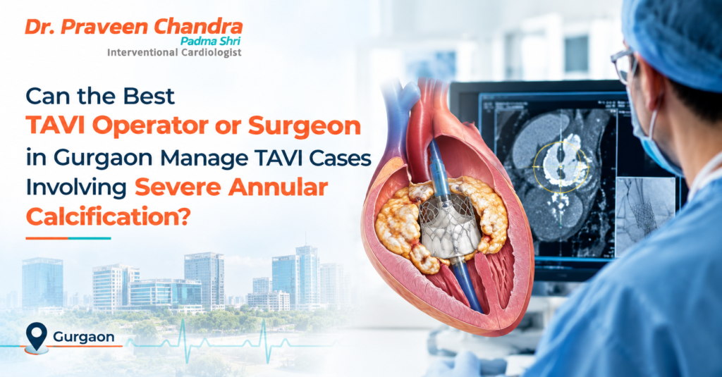
Calcium build-up is the main reason the aortic valve becomes narrow. For most patients with aortic stenosis, some calcium is expected. But when the calcium is severe and extends into the valve ring (the annulus) or the area just below it, TAVI (transcatheter aortic valve implantation) becomes more technically demanding. Patients searching for the best TAVI operator in Gurgaon for this situation are asking an important question: can this be done safely, and what does the team do differently?
This article explains why annular calcification matters, what risks it adds, how experienced teams plan for it, and what to ask before you decide.
Key takeaways
• Severe calcification does not automatically rule out TAVI, but it raises specific risks that need planning.
• CT scanning shows how much calcium there is and exactly where, which strongly influences the plan.
• Valve choice, sizing and balloon use are adjusted to the calcium pattern.
• Surgical backup and emergency readiness are essential in these cases.
• In some patients, surgery or a conservative approach may be the better option.
What is annular calcification, and why does it matter?
The annulus is the ring of tissue where the aortic valve is anchored. In TAVI, the new valve is expanded against the old, calcified valve and the annulus, and it relies on that surrounding tissue to sit securely and seal.
When calcium is heavy or unevenly distributed, particularly in the annulus or the outflow area just beneath the valve, it can:
• Prevent the new valve from expanding fully and evenly
• Leave gaps through which blood leaks back around the valve (paravalvular leak)
• Put extra stress on the surrounding tissue as the valve expands
• Press on the heart’s electrical conduction system, raising the chance of needing a pacemaker
• Increase the chance of calcium fragments travelling to the brain
Risks the team needs to plan for
Risk | Why calcium increases it | How teams plan for it |
Annular or aortic root injury (rupture) | Heavy calcium just below the valve, together with a new valve that is too large for the annulus, can overstress the tissue. This is uncommon but serious. | Careful CT sizing, avoiding oversizing, cautious use of balloons, and surgical and emergency backup on standby. |
Leakage around the valve | Uneven calcium can stop the valve sealing against the wall. | Choosing a valve and size suited to the calcium pattern, and checking the result by imaging during the procedure. |
Need for a pacemaker | Calcium near the electrical pathway can be compressed by the new valve. | Planning the implant depth, and monitoring rhythm closely afterward. |
Stroke | Calcium debris can break off during the procedure. | Minimising manipulation, and using a cerebral protection device in selected cases. |
Coronary artery blockage | Bulky calcified leaflets can be pushed toward the coronary openings. | Measuring heights and leaflet size on CT, and protecting the artery where risk is high. |
How an experienced team approaches severe calcification
1. Measuring calcium on CT
A dedicated cardiac CT scan shows how much calcium is present and where it sits: on the valve leaflets, in the annulus, or in the outflow area below the valve. Location matters as much as amount. Calcium just below the valve is generally considered to carry a higher risk of tissue injury than calcium confined to the leaflets.
2. Choosing the right valve and size
Different valve designs expand and exert force on the annulus in different ways. In heavily calcified annuli, the team considers the design of the valve, how much it is sized relative to the annulus, and how the calcium is distributed. Selecting a size that fits securely without excessive oversizing is a key part of reducing risk. There is no single right valve for every patient, and the choice is individualised.
3. Using balloons carefully
Balloons may be used before the valve is placed, to prepare the narrowed valve, or afterward, to improve expansion. In severe calcification, each use is a judgement call. More aggressive balloon expansion can improve valve fit but may also raise the risk of tissue injury, so the team balances the two based on the patient’s CT findings and on how the procedure is going.
4. Protecting the brain and coronary arteries
When the risk of debris or coronary blockage is high, the team may use a device to catch debris travelling toward the brain, or take steps in advance to protect a coronary artery. These measures are used selectively, not in every case.
5. Being ready for complications
Because the stakes are higher, the setting matters. Cases with severe annular calcification are best performed in a centre with a hybrid cath lab or operating room, a cardiac surgeon available on-site, emergency equipment and a trained team ready to respond immediately. Preparation and rehearsal of the emergency plan are part of good practice.
6. Close monitoring after the procedure
After TAVI, patients are monitored for rhythm disturbances, bleeding, kidney function and valve performance. An echocardiogram checks that the valve is working well and that there is no significant leak. Heavily calcified cases may need closer follow-up of the heart rhythm.
When is surgery or another approach better?
TAVI is not right for every patient with severe calcification. Surgery allows the surgeon to remove calcium directly and fit a valve under direct vision, and may be preferred in younger, fitter patients or when calcification is extreme. In frail or high-risk patients, TAVI may still be the safer choice despite the added complexity. This is exactly the type of decision a heart team should make together.
Questions to ask your TAVI team
• How much calcium do I have, and where is it located on my CT?
• What specific risks does this create for me?
• Which valve and size are you recommending, and why?
• Would surgery be a better option in my case?
• What emergency and surgical support will be available during the procedure?
• How often does your team treat patients with similar calcification?
• What follow-up will I need, especially for heart rhythm?
About Dr. Praveen Chandra
Dr. Praveen Chandra is Chairman of Interventional Cardiology at Medanta Medicity, Gurgaon. He was awarded the Padma Shri by the President of India in 2016 and performed the first percutaneous aortic valve implantation without surgery in India. He has authored over 500 articles and is a reviewer for leading medical journals. His work includes structural heart interventions such as percutaneous tricuspid valve procedures and leadless pacemakers.
When should you see a cardiologist?
Consult a cardiologist promptly if you have aortic stenosis and notice breathlessness on exertion, chest pain, dizziness, fainting or unusual tiredness. Seek emergency care immediately for severe chest pain, sudden fainting or sudden severe breathlessness.
Frequently asked questions
Can TAVI be done with severe calcification of the aortic valve?
In many patients, yes. Calcium is present in most people who need TAVI. However, severe calcification around the valve ring or just below it increases certain risks, so careful CT planning and an experienced team are important.
What is annular rupture in TAVI?
Annular rupture is a rare but serious complication in which the tissue around the aortic valve ring tears during valve implantation. Heavy calcium just below the valve and oversizing of the new valve are among the factors that can raise the risk.
How does the team reduce risks in a heavily calcified valve?
Steps include detailed CT assessment of calcium, choosing a suitable valve type and size, cautious balloon use, protection against stroke where appropriate, and having surgical and emergency support ready.
Is surgery safer than TAVI for severe calcification?
It depends on the patient. Surgery allows direct removal of calcium but carries its own risks, especially in older or frail patients. A heart team weighs both options for each individual.
How do I book a consultation for a heavily calcified aortic valve?
You can book an appointment with Dr. Praveen Chandra’s team by calling +91 99588 72855 or +91 97172 89363.