
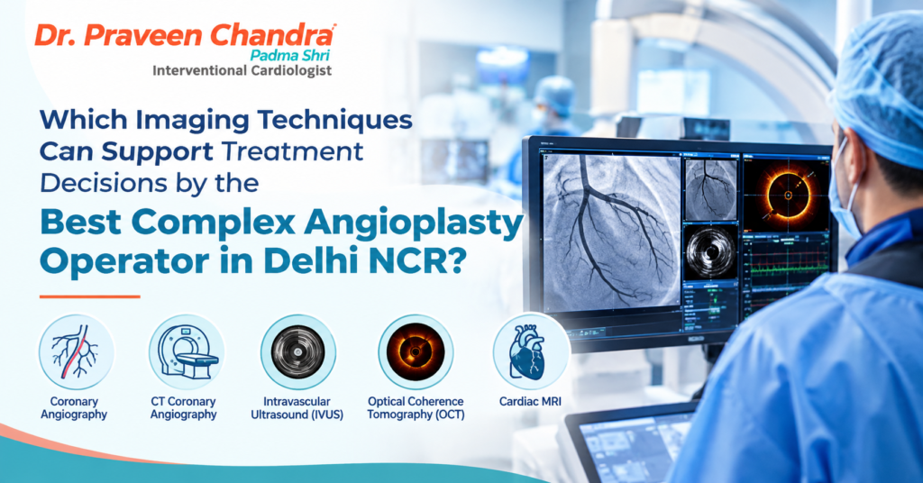
A complex angioplasty needs proper diagnosis, planning, and knowledge of coronary artery disease. In case a person has a blocked heart vessel, atherosclerotic coronary arteries, or an artery within the stent that is narrowed, standard imaging will not be able to provide all the information necessary for planning treatment. Advanced coronary imaging would prove helpful to cardiologists in this regard. For those people searching for the Best Complex Angioplast Operator in Delhi NCR, knowing the importance of coronary imaging would prove beneficial for them.
A person interested in interventional cardiology treatment of coronary artery disease can think of Dr. Praveen Chandra. Imaging does not only show whether there is a blockage. With the help of imaging, a doctor can determine the size of an artery, study the accumulation of plaque, see how a blockage affects the function of an artery, and evaluate whether a stent has expanded. These factors are especially important when dealing with complicated cases.
Why Advanced Imaging Matters for the Best Complex Angioplasty Operator in Delhi NCR
A person gets coronary artery disease due to the formation of a blockage in the arteries that supply oxygen-filled blood to the heart. Due to this blockage in the arteries, there is an obstruction in blood flow to the heart, and one experiences chest pain, difficulty breathing, and even less stamina for exercise. A coronary angiogram is performed to assess these narrow passages. In this procedure, contrast dye is injected into the arteries of the heart, and then the images of X-ray are taken.
This may be relevant when performing angioplasty on a complicated patient. It is possible that two blockages will look identical on an angiogram, yet one will have more calcium or a different impact on blood flow than the other. In this case, while one blockage will be easily treated using stents, the other will require a bit more preparation. This is where advanced imaging techniques become useful, as they give the cardiologist more details regarding the condition of the artery and blockage. Still, this technique is not always needed.
Intravascular Ultrasound (IVUS): Examining the Artery from Within
Intravascular ultrasound is better referred to as IVUS and makes use of ultrasound waves in taking the picture from within the coronary artery. The ultrasound device fitted at the tip of the catheter is inserted into the blood vessel and takes cross-sectional pictures of the artery and the tissues around it. With IVUS, details of the artery such as the artery size, plaques, and calcium deposits are made available. The technique enables the cardiologist to know the true dimensions of the artery and not just the way the artery appears in the angiography pictures.
Furthermore, it can be used post-stenting to see if the stent has expanded adequately and has been properly placed. Should there be a problem with any of those factors, then the operator may think of other ways of treating the problem during the procedure. The primary benefit of IVUS is that it offers important information regarding the artery wall which might not be as clear on traditional angiography.
Optical Coherence Tomography (OCT): High-Resolution Coronary Imaging
Another intravascular imaging modality employed in some of the coronary interventions is optical coherence tomography (OCT). This procedure involves the use of near-infrared light for producing high-resolution images of the internal structures of coronary arteries. OCT can help evaluate the characteristics of the plaque, analyze the interaction of the stent with the vessel wall, and assess other issues that might influence the final result. The detailed pictures produced by OCT are especially helpful when the cardiologist requires more information about a complicated blockage or treated artery. OCT can show that the stent was not completely deployed, that there are some gaps between the stent and the vessel wall, or that there is some protruding tissue between the stent struts.
But OCT has some limitations. It usually needs the injection of a contrast agent to remove blood from the field of imaging. The need for this can affect the suitability of this test in cases where the patient is suffering from poor kidney function and other clinical issues. Both IVUS and OCT provide information about the artery internally, but they cannot be used interchangeably in all situations. The choice of the technique depends on the cardiologist.
Fractional Flow Reserve (FFR): Measuring the Effect of a Blockage
All constrictions observed on coronary angiography are not necessarily associated with a reduction of blood flow to the myocardium. In some cases, there can be an obstruction that is quite apparent but does not really affect the flow of blood to the myocardium, while there can be another constriction that has a more limited appearance but causes blood flow to be reduced. The term fractional flow reserve (FFR) is used for this purpose.
The results help the cardiologist determine how appropriate it would be to perform any form of treatment for the blockage. FFR proves to be most effective in cases where there are intermediate-level blockages found on angioscopy and it is difficult to ascertain if stenting needs to be done or not. In such cases, rather than taking only the appearance of the arteries into account, the doctor uses the results of pressure measurement to make a conclusion. But then again, the results are to be combined with other clinical factors too.
Instantaneous Wave-Free Ratio (iFR): An Alternative for Functional Assessment
The third technique used to test the significance of coronary narrowing is called the instantaneous wave-free ratio, which is commonly referred to as iFR. As opposed to FFR, iFR involves the use of a wire that can measure pressure across the narrowing, but at a particular part of the cardiac cycle of the heart. The measurement is not usually carried out using drugs to increase coronary blood flow. This makes it a convenient way of testing in some cases. It is also useful for patients who do not wish to be given the drugs in the usual FFR test.
FFR and iFR both assist in finding whether the narrowing of the vessel causes an impact on the blood flow. The results of the research are useful in determining whether there should be more angioplasty, drug treatment, or examination needed. It is important to remember that neither of these tests actually visualizes the structure of the vessel wall.
How Imaging Helps Treat Heavily Calcified Blockages
The excessive calcification of the coronary arteries makes the process challenging. The occurrence of calcium may not allow the balloon to inflate completely and may influence the inflation of a stent. If the stent fails to inflate, this can cause different complications. IVUS is beneficial in helping the cardiologist to ascertain the existence and severity of the calcium deposits in the arteries. This is important in helping the cardiologist decide how to prepare the blockage when placing the stent. Depending on the nature of the anatomy and the presentation of the patient, the procedure of choice can be rotational atherectomy and intravascular lithotripsy.
But this cannot be done in all cases. It all depends on the type of blockage, the imaging, and the assessment by the specialist. In some cases, imaging can even be done post-procedure to evaluate how well the stents have been placed, and whether there are any issues that require correction. This is more so in those cases where it becomes difficult to assess the outcome just through angiography. If you are consulting a complex angioplasty operator in Delhi NCR, you should try to find out if imaging would help in your case.
Comparing Different Imaging and Assessment Techniques
Technique | Main purpose | Role in treatment planning |
Coronary angiography | Shows the outline of coronary arteries | Identifies the location and apparent severity of blockages |
IVUS | Examines the artery wall and plaque from within | Helps assess vessel size, calcium, and stent expansion |
OCT | Produces detailed images of inner artery structures | Helps evaluate plaque details and stent positioning |
FFR | Measures pressure differences across a blockage | Helps determine whether narrowing restricts blood flow |
iFR | Assesses the functional effect of a blockage | Supports decisions about whether selected blockages need intervention |
Supports decisions about whether selected blockages need intervention
It all depends on the nature of the clinical question being asked. Angiography determines the location of the problem, while IVUS and OCT give the structural details; FFR and iFR are functional determinants. Sometimes, one modality is enough to determine the answer, while at other times, a combination is needed.
How Imaging Supports Better Stent Placement
Success of angioplasty goes beyond mere widening of the narrowed artery. The stent must be of a suitable size and correctly positioned with respect to its point of beginning and ending so that blood can pass through the widened segment of the artery. In some complicated cases, intravascular imaging may aid the physician in assessing the appropriate stent size and the location at which stenting should be begun and finished. It can be particularly useful for those cases when there is an elongated plaque, irregular diameter of the artery, and difficulties in assessment of coronary anatomy. Once the stent is placed, IVUS or OCT can help the cardiologist to evaluate whether there are any problems such as improper stent expansion or lack of stent-vessel contact.
Imaging may be helpful if symptoms appear after the angioplasty procedure. Imaging may assist in finding out if the reason for that is the development of tissue within the stent, lack of dilation, or another structure-related cause. While imaging helps to have more data at hand for the operator, it does not guarantee the lack of complications during the procedure. Long-term results depend on the degree of coronary artery disease and other factors.
Choosing the Right Cardiologist for Complex Angioplasty
Patients who have multiple stenoses or complicated coronary anatomy often have some questions concerning their treatment. The patient may wonder whether angioplasty, bypass surgery, or drugs are needed for his/her disease. The discussion will help the cardiologist understand the patient’s symptoms, medical history, angiography results, heart function, and other information and provide recommendations about the treatment. When choosing the specialist, the patient should consider the experience in performing complex coronary procedures, the approach to risk assessment, and the ability to explain the treatment techniques to the patient. It is also possible to ask whether IVUS, OCT, FFR, or iFR are needed in this particular case.
Conclusion
Imaging tools play an important part in certain complex angioplasty cases. IVUS and OCT give a comprehensive view of the coronary artery and stent, while FFR and iFR help to evaluate the impact of a blockage on blood flow. They can be used to make choices about lesion preparation, stent placement, and treatment options. Selection of the Best Complex Angioplasty Operator in Delhi NCR requires knowledge of the doctor’s expertise, complexity of the procedure, and reasoning behind the suggested treatment. Dr. Praveen Chandra can be approached by patients seeking an interventional cardiology consultation for coronary artery disease. It is important for a personalized assessment to be done before knowing which of the above treatments would be the best for their disease.
Frequently Asked Questions
1. Is advanced imaging essential for every angioplasty?
No. Many operations can be done using only coronary angiography. The use of IVUS and OCT will be more appropriate in certain cases when there is a need for additional information regarding the wall of the artery, calcium deposits, or stent expansion.
2. What is the difference between IVUS and OCT?
IVUS creates images from inside the artery with the use of ultrasound waves, whereas OCT creates images of the inner layers of the vessel using near-infrared light. The cardiologist decides which method should be used depending on the patient’s case and the information required.
3. Can FFR and iFR help in avoiding unnecessary stent placement?
Yes. These methods can assist in determining the degree to which a blockage reduces the flow of blood.
4. What Imaging Technique Should Be Used for Calcified Coronary Lesions?
There is no ideal technique for all patients. IVUS and OCT imaging can be used to evaluate the calcium content and plan the procedure in selected cases. The right imaging technique and plaque preparation procedure should be based on the individual blockage and the condition of the patient.
5. How Can I Find the Complex Angioplasty Operator in Delhi NCR?
You need to find a cardiologist with proper experience in complex coronary procedures, conservative treatment planning, and the ability to explain the options to the patient. The doctor should discuss the pros and cons of angioplasty, the need for imaging techniques, and alternatives to it.